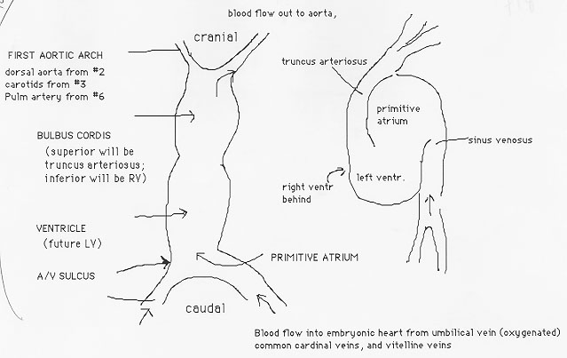
The heart forms from cords of cardiogenic mesoderm
primitive heart tube
Þ fusion of endothelial heart tubes
the paired dorsal aortae develop and connect with endocardial tubes before folding begins
Þ form 1st aortic arch
3 pairs of vessels supply inflow to heart:
Common cardinal veins (venous blood from embryo body)
Vitelline veins (blood from yolk sac)
Umbilical veins (oxygenated blood from placenta)
During week 5-8, heart tube undergoes folding and remodeling
(see figure below)
develops bulges and sulci
Þ primordial heart chambers
Inferior (inflow end)
Þ sinus venosus – common cardinal veins drain here; primitive atrium; primitive ventricles – future left ventricle; bulbis cordis – future right ventricle, outflow regions of both ventricles
all systemic venous blood drains into right atrium
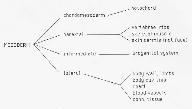
splanchnic mesoderm
Þ primitive myocardium
Circulation Changes
Food absorption
Þ yolk sac or placenta; Lungs are not used
5th – 6th week (Fig 3)
septum primum and septum secundum separate right and left atria
Þ allows right to left shunting in heart atria; foramen ovale (shunts partially oxygenated blood to the body, with less blood forced to the lungs); septa fuse ~2 – 3 months of life
Vasculature
Blood vessels begin in the extra-embryonic mesoderm covering the yolk sac, connecting stalk and chorion
Embryonic blood vessels develop ~2 days later
Blood vessel formation (mesodermal) – do not grow like trees, grow in segments which become connected
angioblasts form blood islands
Þ cavities
angioblasts flatten, form endothelial cells that line the cavities