Definition: a motor system that innervates the heart, smooth muscle, and glands. Sensory is NOT part of the system
Autonomic pathway organization:
primary visceral sensory neuron in peripheral ganglia Þ secondary order visceral sensory relay that projects locally and to the thalamus Þ visceral motor neurons. All autonomic cell groups are regulated by neurons above the level of the spinal cord and brainstem
Basic unit:
two-neuron chain that consists of:
Preganglionic – cell body is located in the gray matter of the spinal cord or brainstem. Axon projects out of the CNS onto the postganglionic neuron
Postganglionic – cell body is located in a ganglion outside the CNS. Axon projects onto an end organ (heart, smooth muscle, or gland).
Central and reflex control:
the basic two neuron autonomic effector does not operate autonomously. Activity of the preganglionic neuron is modulated reflexively and/or by specific centers in the brain (i.e. medulla, pons, hypothalamus, cerebral cortex)
3 divisions
: Sympathetic, Parasympathetic, and Enteric
Anatomy
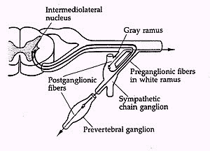
Sympathetic (thorcolumbar) nervous system
Preganlionic neuron – cell body in the intermediolateral cell column of spinal cord segments T1 to L2,3. Axons are myelinated (termed white rami), pass out of the spinal ventral root, enter chain root ganglia and synapse with postganglionic neurons (connections converge and diverge) in ganglia of peripheral nervous system
Postganglionic neuron
– unmyelinated; innervate heart, smooth muscle, and glands. Emerge as gray rami innervates blood vessels (vasomotor), hair (pilomotor), and sweat glands (sudomotor). Cell bodies located in 2 types of ganglia:
(1) Chain ganglia
(vertebral paravertebral ganglia) – 2 chains located next to each vertebrae on both sides. Each chain has 1 ganglion except cervical region which supply symp innervation to the heart and head (cervical ganglia fusion of superior, middle, and inferior cervical ganglion)
Stellate ganglion
– fusion of inferior and 1st thoracic ganglion
Sympathetic trunk
– each chain ganglion and connecting axons (there are 2 – one on either side of spine)
(2) Prevertebral ganglia
(collateral ganglia): irregular ganglionic mases that surround visceral branches of aorta. Innervate abdominal and pelvic viscera (stomach, intestine, adrenal medulla, bladder). Paired or unpaired. Names based on arteries with which they are associated (celiac ganglion, superior mesenteric ganglion, and inferior mesenteric ganglion)
Parasympathetic (craniosacral) nervous system
Preganglionic: Associated with cranial nerves: III (oculomotor), VII (facial), IX (glossopharyngeal), X (Vagus)
Innervates smooth muscle and glands of head, heart, and abdomen
X (Vagus) is most important, since it supplies all thoracic and abdominal viscera; motorneurons are found in dorsal motor nucleus (contains parasympathetic only) and nucleus ambiguus.
Unmyelinated and long (synapse with ganglia close to end organ, in contrast to sympathetic, which have chain ganglia)
Sacral location: intermediolateral gray segments S2-S4 Þ form the pelvic nerve innervates pelvic viscera
Postganglionic:
short and unmyelinated (cell body in ganglia next to end organ), Cranial ganglia (ciliary, otic, submaxillary)
Sensory Inputs:
Afferent innervation from primary sensory neurons in cranial and dorsal root ganglia (DRG)
– receives primary afferent projections from visceral sensory neurons in cranial ganglia
Control of Respiration and Blood Pressure: primary sensory neuron Þ nTS (interneuron) Þ spinal motor neuron
Deep Breathing Test
Þ parasympathetic
Axon Reflex Sweat Test
Þ post-ganglionic sympathetic
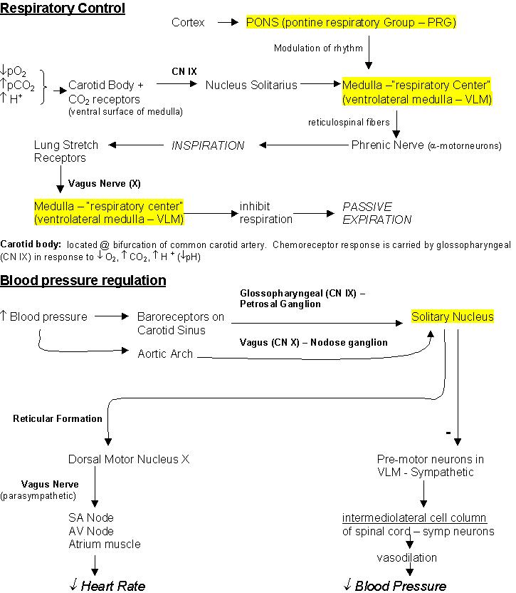
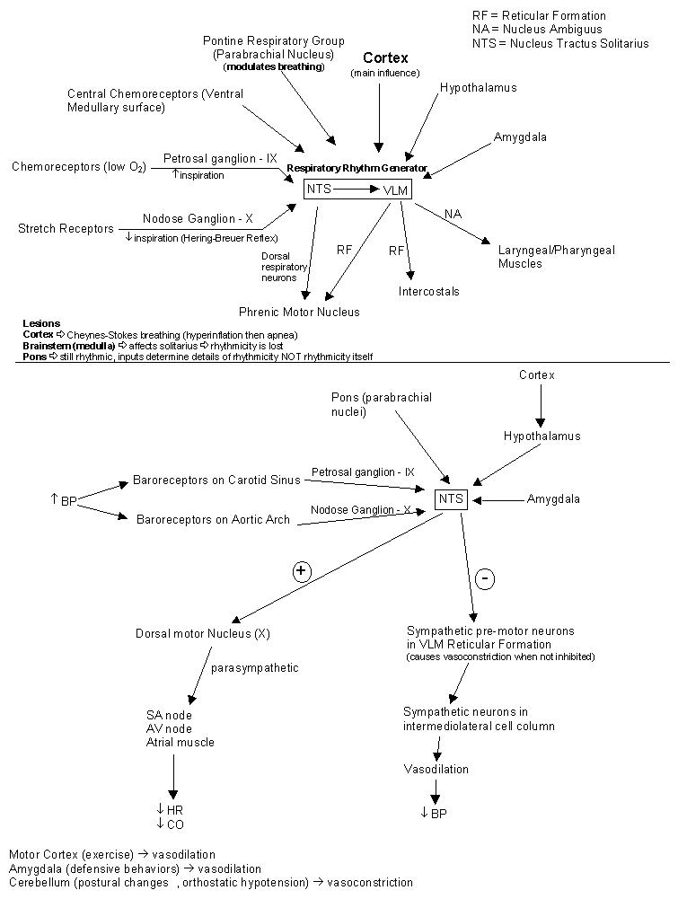
Respiratory Control
Respiratory rhythm generation
: generation of breathing patterns is Autonomous; NO sensory input is required
Brainstem neurons involved:
(1) Nucleus Tract Solitarius
– contains (1) Afferent chemo and pulmonary stretch receptors (2) dorsal respiratory neurons (fire during inspiration) that synapse in phrenic motor nucleus Þ innervates diaphragm
(2) Ventrolateral Medulla (VLM):
contain pacemaker cells of respiration Þ generate bursts of spikes that are the source of the respiratory rhythm
vagal motorneurons in dorsal nucleus ambiguus (innervate laryngeal and pharyngeal muscles)
ventral lateral reticular formation (RF) neurons – innervate intercostal and phrenic motorneurons in spinal cord
Dorsolateral pons (pontine respiratory group, PRG) – modulate activity of the respiratory rhythm generator;
PRG has reciprocal connections with ventrolateral reticular formation, nTS, limbic system (amygdala), hypothalamus
PCO2 – sensed within the CNS (ventral surface of medulla) connected to nucleus solitarius
Lesions
Cortex
Þ Cheynes-Stokes breathing (hyperinflation then apnea) Abnormal but rhythmic why? Brainstem is intact medullary regulator is still operating
Brainstem (medulla)
Þ affects solitarius Þ rhythmicity is lost
Pons
Þ still rhythmic, inputs determine details of rhythmicity NOT rhythmicity itself
Blood Pressure Regulation
Determinants of BP: Cardiac output, peripheral resistance (vascular tone)
Cardiac output:
regulated by sympathetic and parasympathetic (vagal)
Vascular tone:
maintained by tonic vasomotor activity of sympathetic preganglionic neurons (SPN) in the intermediolateral cell column (ILC) of thoracic and lumbar spineal cord.
Ventrolateral medullary reticular formation
(pre-motor neurons) – tonic input from this formation project to interomedial column of sympathetic chain and maintains SPN fibers (clonadine stimulates this area; close to ventral respiratory group)
Brain stem neurons involved:
(1) nTS
– site of afferent baro- and chemoreceptors, secondary neurons: homeostasis primarily attributable to baroreceptor reflex
sensory fibers from aortic arch (via nodose ganglion X) and carotid sinus (via petrosal ganglion IX) baroreceptors terminate in specific nTS subnulei
afferent stimulation causes hypotension and bradycardea; lesion of nTS
Þ hypertension and abolition of baroreflexes
nTS neurons excite cardiac vagal motorneurons
Þ ß HR and CO
baroreflex has inhibitory component; nTS neurons make inhibitory connections with symp pre-motor neurons in VLM
(2) VLM
– pre-motor neurons to symp neurons in the ILC
Supramedullary structures
(hypothalamus, cerebellum, cortex) – mediate cardiovascular adjustments to changing behavioral demands (i.e. stress, etc), but structures above medulla cannot maintain BP
Vasodilation pathways: arise in motor cortex (used during exercise) and amygdala (defensive behaviors)
Vasoconstriction pathways: cerebellum – mediates homeostasis of BP during postural changes (e.g. orthostatic hypotension)