MRI Landmarks
MRI – T1 and T2
– Spin density
- Recall that the MRI puts the patient in a magnetic field that aligns all of their hydrogen atoms so that they are all spinning on one particular axis. A radio frequency pulse is than delivered to knock these atoms out of alignment. When these atoms return to the alignment set by the magnetic field, they send out a frequency pulse picked up by a coil detector which are then processed to produce an image. Since different tissues have different densities of hydrogen atoms, this can be used to discriminate tissue.
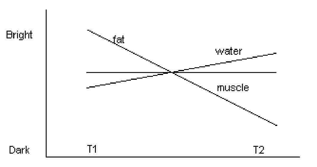
- The "T" refers to the 2 different spin axes of the H atom (it spins and gyrates). When they are knocked out of alignment, The H atoms return to the axis set by the MRI on these 2 different axes at different speeds. Based on these 2 different types of spin, one tissue type can deliver 2 different types of frequency to the detector. By this, you can represent one tissue with different intensities on the MRI image. So, on a T1 weighted image fat shows up brightly and on a T2 weighted image water shows up brightly. Muscle can be delineated on both weighted images but does not light up.
Radiologic Anatomy
Thalamus – 2 egg shaped structures right below the corpus callosum
- Massa Intermedia
(aka interthalamic adhesion) – connection of the thalamus on the midline
Mammilary bodies – little egg structures right below the hypothalamus; make up the rostral end of the fornix.
Fornix – connects the hippocampus to the hypothalamus; 3 parts:
- (1) columns
(2)
- (2) body
– connected by a commissure or hippocampal commisure
- (3)
crura (legs)
- the body hangs off the corpus callosum in the midline by the septum pellucidum
Midbrain (mesencephalon) sagittal view Þ inferior to the lateral ventricles and superior to the pons
- transverse cut Þ Peduncles (cerebral crus: contains the pyramidal tract (the mickey mouse ears))
- Internal Capsule
– superior continuation of the cerebral crus, separates the thalamus from the caudate and lentiform nuclei; major connection of cortex to the brain stem and spinal cord
- Coronal radiata –
superior and lateral continuation of internal capsule, major part of cerebral hemisphere’s white matter
Tectum (quadrigeminal plate) – "roof" of the midbrain; dorsal part of the midbrain that contains superior and inferior colliculi; the cerebral aqueduct (duct of Sylvius) runs through the posterior body of the midbrain
Tegmentum – main part of the substance of the midbrain; transverse cut runs from the substantia nigra to the cerebral aqueduct
Corpus callosum – the great commissure (connecting) plate of nerve fibers interconnecting the cerebral hemispheres
- 4 parts (from rostral to caudal): rostrum, genu (knee), trunk (a.k.a. body), splenium
Grey matter ribbon – outer cortex of the frontal lobe on coronal section; this is a descriptive term that indicates the frontal lobe is showing early signs of edema from an infarct or encephalitis
Sylvian Fissure (lateral cerebral sulcus) – deepest, most prominent fissure in the brain that separates the frontal from the temporal lobe; branches of the middle cerebral artery run within its course
- Insula
– (Island of Reil) – On coronal section – oval region of white matter over the external capsule
Ventricles – 2 lateral ventricles, each of which has 4 parts: frontal horn (anterior), central part, temporal (inferior) and occipital (posterior) horn
3rd Ventricle is connected to 2 lateral ventricles via Interventricular foramen (of Monroe)
4th ventricle connects with the 3rd via the cerebral aqueduct (of Sylvius)
4th ventricle has a lateral outlet (foramen of Luschka) and a median aperture (foramen of Magendie)
Cistern – dilation of the subarachnoid space that contains CSF – 4 cisterns:
- (1)
interpeduncular – in front of the pons
- (2) prepontine –
below the Pons
- (3) chiasmatic
– in front of infundibulum
- (4) quadrigeminal
– above tectum
Cerebellum – large posterior brain mass lying over the pons. Divided in midline by gray matter (Vermis)
- Vermis
(worm) – superior and inferior sections; connects the two hemispheres of the cerebellum